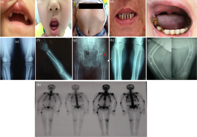
Figure 1. The clinic characteristics and radiographic signatures of the HPP patients.
(a,b) Early deciduous teeth loss of FM1-1 and FM2-1. (a) FM1-1 had only two maxillary canines left at 6 years of age; (b) FM2-1 had only eight primary teeth left at 8 years of age. The permanent teeth present were all molars. (c) Pectus excavatum of FM2-1; (d) sparse teeth of FM2-2; (e) radiographs of both knees of FM2-2 showed slight skeletal hyperostosis; (f) radiographic examination of FM3-1 showed signs of rickets in the distal ulna and radius; (g) the anteroposterior of the pelvis of FM3-1 showed subluxation of the bilateral hip (red arrow) and calcium deposition adjacent to the great trochanter of the left femur (white arrow); (h) X-ray examination of FM4-1demonstrated cortical thickening in fibula and tibia bone; (i) early deciduous teeth loss of FM5-1. The permanent teeth present were all lower incisors and lower molars, and the lower molars displayed hypocalcified enamel; (j) fracture lines of humerus and femur of FM5-1; (k) bone scan of FM5-1 showed multiple areas of increased tracer uptake in the skull, ribs, and femurs.

