
Figure 5:
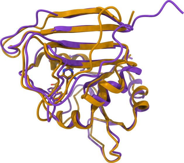
Representative example of a structure model constructed by eThread. The model of DHFR (purple ribbons) complexed with BDBM50329610 is superposed onto the crystal structure of homologous DHFR from S. aureus (gold ribbons) complexed with UCP1106. Ligands bound to target proteins are shown as solid sticks (BDBM50329610 is purple and UCP1106 is gold) with non-carbon atoms colored by atom type (O—red, N—blue).