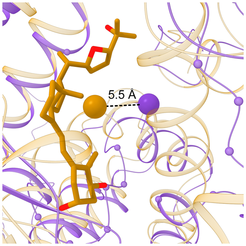
Figure 7:

Representative example of a binding site detected by eFindSite. The model of VDR (purple ribbons) is superposed onto the crystal structure of homologous VDR from human (gold ribbons) complexed with a synthetic analog of vitamin D (gold and red sticks). Cα atoms of binding residues predicted in the VDR model by eFindSite are shown as small spheres. Large spheres connected by a dashed black line are placed at the location of the predicted pocket center (purple) and the geometric center of vitamin D analog (gold).
