Figure 1.
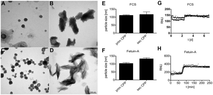
Calciprotein particle CPP formation in protein solutions supersaturated with calcium and phosphate. (A,B,E,G) CPP were prepared in DMEM supplemented with 1.0 mM calcium (total Ca 3.8 mM), 3.5 mM phosphate (total Pi 4.4 mM), and 10% fetal calf serum FCS. (C, D,F,H) Alternatively, CPP were prepared in HEPES/NaCl buffer supplemented with 10 mM calcium, 6 mM phosphate, and 1.0 mg/ml purified fetuin-A protein. (A–D) CPP were harvested by centrifugation and analyzed by electron microscopy or by nanoparticle tracking analysis (E,F). (A–D) Depict representative CPP in their primary (A,C) and secondary (B,D) state. DMEM/FCS-derived and HEPES/fetuin-A-derived primary CPP were similar, but DMEM/FCS-derived secondary CPP appeared denser, yet less crystalline because of higher protein content. Electron microscopy shows that primary and secondary CPP are distinct in shape and size, while nanoparticle tracking analysis (E,F) indicated similar particle sizes of around 100–125 nm for all preparations. Continuous nephelometer measurements (G,H) indicated a sharp transformation of primary into secondary CPP around 2.5 days and 75 min in the presence of DMEM/FCS and HEPES/fetuin-A protein, respectively.

