
Figure 4.
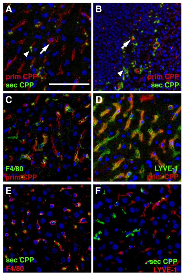
Differential clearance of primary and secondary CPP. Mice were injected with a mixture of fluorescence labeled primary (red) and secondary CPP (green) and the major clearance organs liver (A,C–F) and spleen (B) were harvested 10 min after injection, sectioned and analyzed for the presence of CPP. Primary CPP (prim CPP, arrows in A,B) and secondary CPP (sec CPP, arrow heads in A,B) showed distinct non-overlapping distribution in liver (A), and spleen (B). (C–F) Co-localization with the macrophage-specific marker F4/80 and the liver sinusoidal endothelial LSEC-specific marker LYVE-1 suggested that primary CPP were predominantly cleared by LYVE-1-positive LSEC, and secondary CPP were predominantly cleared by F4/80-positive liver Kupffer cell macrophages. Scale bar: 25 μm.