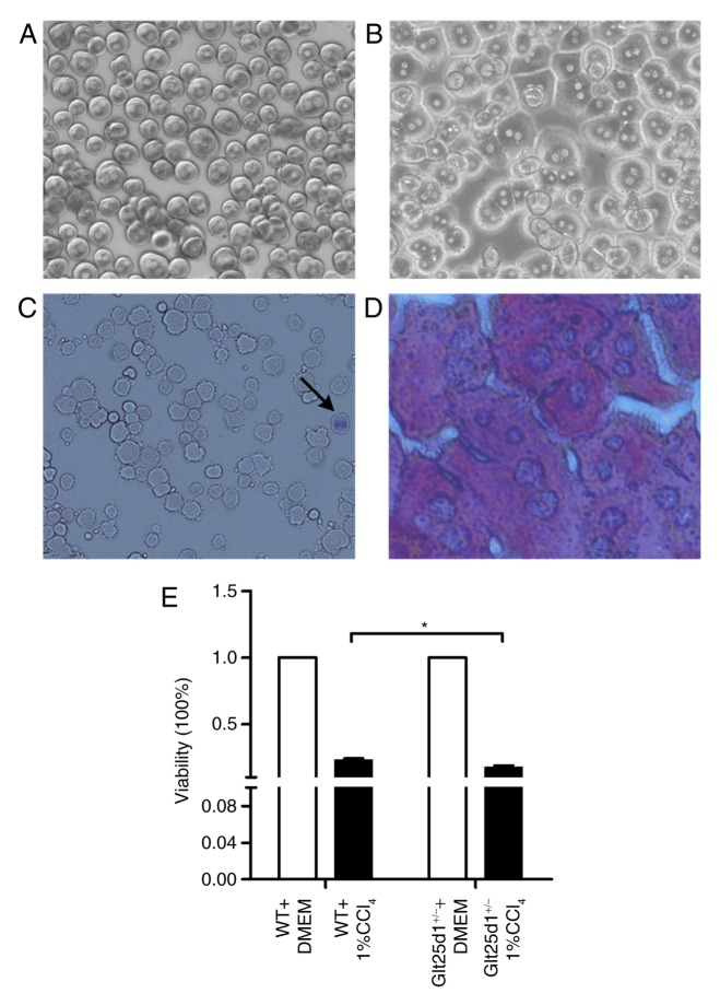
Figure 4.
Glt25d1+/− primary hepatocytes show increased viability loss upon treatment with 1% CCl4. (A) Morphology of mouse primary hepatocytes following initial plating (magnification, ×100). (B) Morphology of mouse primary hepatocytes following incubation for 4 h (magnification, ×100). (C) Viability of primary hepatocytes evaluated by trypan blue exclusion. The arrow indicates a dead cell (magnification, ×100). (D) Purity of primary hepatocytes identified by PAS staining (magnification, ×400). (E) Effects of 1% CCl4 treatment for 24 h in primary hepatocytes. *P<0.05; significance determined by Student's t-test. Glt25d1, collagen β (1-O) galactosyltransferase 1; CCl4, carbon tetrachloride; WT, wild-type.

