Figure 1.
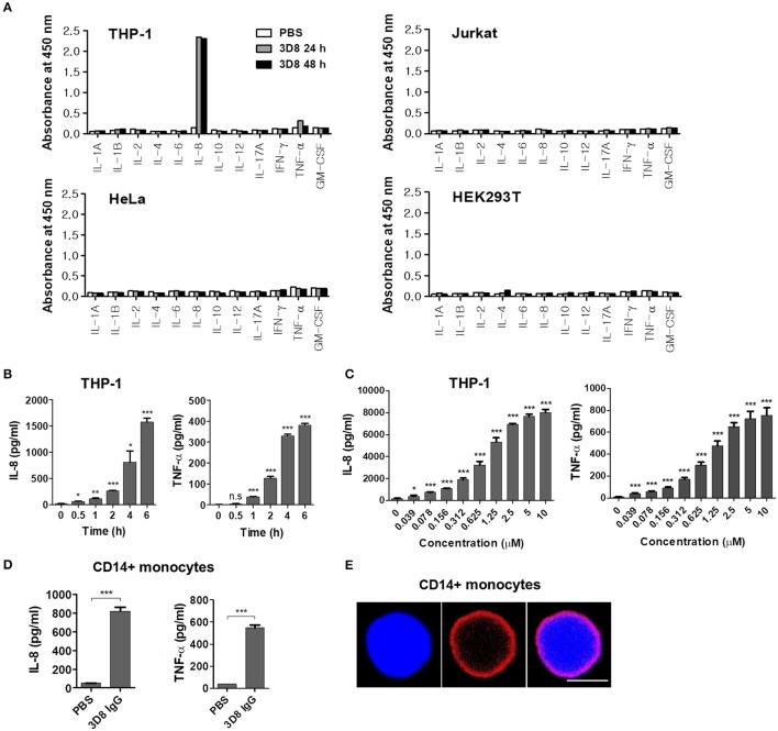
Cytokine secretion by human cells treated with internalizable 3D8 IgG antibodies. (A) Cytokine production by different cell lines. Cells were exposed to 10 μM 3D8 IgG for 24 or 48 h, and cytokine secretion was analyzed (in duplicate) using ELISA kits. (B,C) THP-1 cells were treated with 5 μM 3D8 IgG antibodies for the indicated times at 37°C (B) or with different concentrations of 3D8 antibody for 6 h at 37°C (C). The amount of IL-8 and TNF-α in culture supernatants was analyzed using ELISA kits. (D) Cytokine secretion by human primary CD14+ monocytes exposed to 5 μM 3D8 IgG antibodies. (E) Confocal microscopy. Human primary CD14+ monocytes were exposed to 5 μM 3D8 IgG antibodies for 6 h at 37°C. After fixation and permeabilization, cells were incubated with Dylight 550-conjugated goat anti-human IgG/Fc. Scale bar, 5 μm. Data are expressed as the mean ± standard error of the mean (three independent experiments). All p-values were calculated using a two-tailed Student's t-test (n.s., not significant; p > 0.05; *p < 0.05; **p < 0.01; and ***p < 0.001 vs. negative control).

