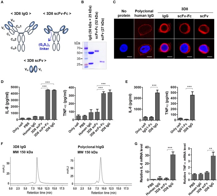
Figure 2.
The Fc region of internalizable 3D8 IgG antibody triggers production of IL-8 and TNF-α by THP-1 cells. (A) Schematic representation of the different 3D8 molecules. (B) SDS-PAGE analysis of 3D8 antibodies purified from the culture supernatant of HEK293F cells transfected with genes encoding the different antibody formats. (C) Confocal microscopic analysis of intracellular localization of 3D8 antibodies. Cells were exposed to 5 μM antibodies for 6 h at 37°C. After fixation and permeabilization, cells were incubated with Dylight 550-conjugated goat anti-human IgG/Fc to detect IgG and scFv-Fc. 3D8 scFv was detected by a polyclonal rabbit anti-3D8 scFv, followed by TRITC-conjugated goat anti-rabbit IgG. (D,E) Cytokine secretion. Cells were exposed to 5 μM 3D8 antibodies, polyclonal human IgGs (D), and 5 μM isotype control (human IgG1-Fc fragment) (E) for 6 h at 37°C. The amount of IL-8 and TNF-α in the culture supernatant was measured using ELISA kits. (F) Size exclusion chromatography of 3D8 IgG and polyclonal human IgG. The indicated molecular weights were interpolated using a standard curve set up using proteins with known mass and retention time. (G) Quantitative RT-PCR of mRNA encoding IL-8 and TNF-α in cells exposed to 5 μM 3D8 antibodies for 6 h at 37°C. (D,E,G) Data are expressed as the mean ± standard error (three independent experiments). All p-values were calculated using a two-tailed Student's t-test (**p < 0.01 and ***p < 0.001).

