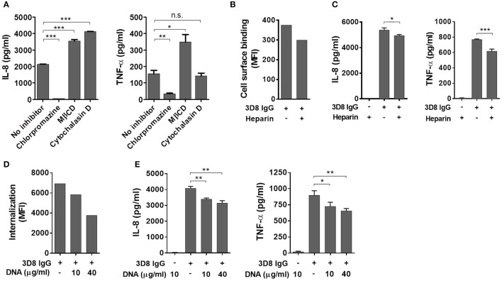
Figure 3.
Induction of inflammatory cytokines by internalized IgG. (A) ELISA. Prior to exposure to 5 μM 3D8 IgG for 6 h, THP-1 cells were treated with endocytosis inhibitors [10 μg/ml chlorpromazine, 5 mM MβCD, or 1 μg/ml cytochalasin (D)]. The amount of IL-8 and TNF-α in the culture supernatant was measured using ELISA kits. (B) Binding of 3D8 IgG (5 μM) to the cell surface in the presence of 10 μg/ml heparin was examined by flow cytometry. (C,E) ELISA. THP-1 cells were treated with a mixture of 3D8 IgG (5 μM) and heparin (10 μg/ml) (C), or with a mixture of 3D8 IgG (5 μM) and the indicated concentrations of DNA (E). The amounts of IL-8 and TNF-α in the culture supernatant were measured using ELISA kits. All p-values were calculated using a two-tailed Student's t test (n.s., not significant; p > 0.05; *p < 0.05; **p < 0.01; and ***p < 0.001, vs. negative control). n.s., not significant. (D) Internalization of 5 μM 3D8 IgG into THP-1 cells in the presence of the indicated concentrations of DNA isolated from THP-1 cells was examined by flow cytometry.

