Figure 4.
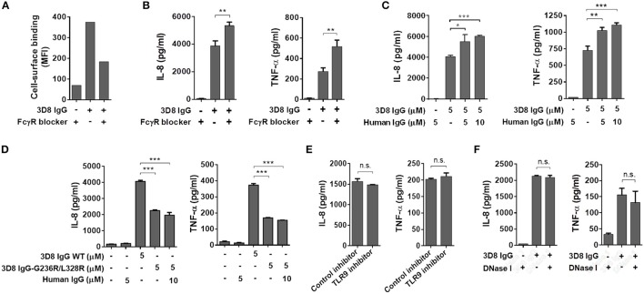
Production of inflammatory cytokines triggered by internalized IgG does not occur via cell surface FcγR- or intracellular TLR9-mediated signaling pathways. (A) Flow cytometry analysis of 3D8 IgG (5 μM) binding to THP-1 cells in the presence of an FcγR blocker (10 μg/ml). (B,C,E,F) ELISA. Prior to exposure to 3D8 IgG (5 μM) for 6 h, THP-1 cells were treated with an FcγR blocker (10 μg/ml) (B), the indicated concentrations of human polyclonal IgG (C), a TLR9 inhibitor (5 μM) (E), or 1 U/ml DNase I (F) for 1 h at 37°C. (D) THP-1 cells were treated for 6 h at 37°C with either 3D8 IgG-G236R/L328R (5 μM) or a mixture of 3D8 IgG-G236R/L328R (5 μM) and polyclonal human IgG (10 μM). The amounts of IL-8 and TNF-α in the culture supernatant were measured using ELISA kits. All p-values were calculated using a two-tailed Student's t-test (n.s., not significant; p > 0.05; *p < 0.05; **p < 0.01; and ***p < 0.001).

