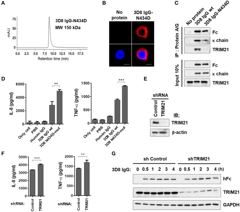
Figure 5.
Internalizable IgG-mediated production of IL-8 and TNF-α is not mediated by TRIM21. (A) Size exclusion chromatography of 3D8 IgG-N434D. (B) Confocal microscopy. THP-1 cells were exposed to the 3D8 IgG-N434D mutant (5 μM) for 6 h at 37°C. After fixation and permeabilization, cells were stained with Dylight 550-conjugated goat anti-human IgG/Fc. (C) Co-immunoprecipitation of 3D8 IgG antibodies and TRIM21. THP-1 cells were exposed to 3D8 IgG antibodies (5 μM) for 6 h at 37°C. Cell lysates were then co-immunoprecipitated with Protein A/G. Samples were then analyzed by immunoblotting with antibodies specific for TRIM21, the Fc portion of human IgG, and human IgG κ light chain. Input controls comprised 10% total cell lysate. (D) ELISA. THP-1 cells were treated with 3D8 IgG antibodies (5 μM) for 6 h at 37°C, and the amounts of IL-8 and TNF-α in the culture supernatants were measured using ELISA kits. (E) Western blot analysis of shRNA-mediated knockdown of TRIM21 in THP-1 cells. (F) ELISA. IL-8 and TNF-α were measured in TRIM21-knockdown THP-1 and control cells exposed to 5 μM 3D8 IgG antibodies for 6 h at 37°C. (D,F) Data are expressed as the mean ± standard error (three independent experiments). All p-values were calculated using a two-tailed Student's t-test. Statistical significance is indicated on the graphs (**p < 0.01 and ***p < 0.001). (G) Immunoblot analysis of TRIM21 and human IgG H chain expression in TRIM21-knockdown THP-1 cells and control cells. GAPDH was used as a loading control.

