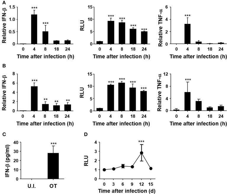
Figure 1.
Induction of type I IFN by O. tsutsugamushi infection. Mouse embryonic fibroblasts (MEFs, A) and bone marrow-derived macrophages (BMDMs, B) were infected with O. tsutsugamushi and harvested at the indicated time points. Total RNA was isolated from the infected cells and the relative levels of IFN-β (left) and TNF-α (right) transcripts, normalized with β-actin mRNA, were determined by qRT-PCR. Type I IFN bioactivity (middle) was also analyzed in L929 cells containing a stable ISRE-luciferase reporter plasmid after stimulation with culture supernatants collected at the indicated times. (C) Secreted IFN-β from infected BMDMs was analyzed by ELISA after 18 h of infection. U.I., uninfected; OT, O. tsutsugamushi-infected. (D) Mice were infected with 5 × LD50 of O. tsutsugamushi and type I IFN bioactivity in plasma collected at the indicated times was determined in L929 cells containing a stable ISRE-luciferase reporter plasmid. Data represent mean ± SD of three independent experiments. Statistical significance was determined by one-way analysis of variance (ANOVA) followed by Newman-Keuls t-test for comparisons with uninfected control. ***p < 0.001, **p < 0.01. RLU, relative luciferase unit.

