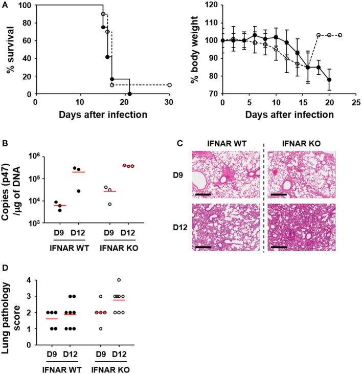
Figure 3.
Effect of type I IFN signaling on mortality, pathogenesis, and bacterial loads in in vivo infection of O. tsutsugamushi. (A) Wild type (black circle) and IFNAR-deficient (white circle) mice (n = 10) were intraperitoneally infected with 5 × LD50 of O. tsutsugamushi and survival rate and weight changes (relative to initial body weight on day 0) were monitored for a month. (B) Bacterial loads in the lungs of infected mice (wild type or IFNAR KO, n = 3/group) were assessed by qRT-PCR using primer sets detecting the p47 gene of O. tsutsugamushi. The infected tissues were collected at the indicated days after infection. (C) Lung tissue sections collected from mice at the indicated days after infection were stained with hematoxylin and eosin and representative scanned images are presented (see also Figure S2). Bar, 300 μm. (D) Pathological scores of infected lungs (D9: n = 5, D12: n = 8) are presented. Red lines, mean.

