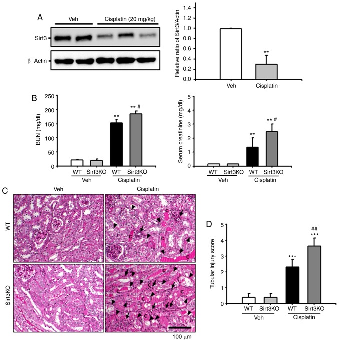
Figure 1.
Effects of Sirt3 on cisplatin-induced renal tubular damage. (A) Sirt3 expression in kidney tissue from WT mice injected with Veh or cisplatin (20 mg/kg body weight) was evaluated by western blotting. Data obtained from densitometric analysis are presented as the relative ratio of each protein to β-actin. The relative ratio of protein measured in kidneys from vehicle-injected mice is arbitrarily presented as 1. **P<0.01 vs. Veh. (B) Kidneys in Sirt3KO and WT mice were harvested 3 days after treatment with cisplatin (20 mg/kg body weight). Blood samples were collected 72 h following cisplatin treatment, and BUN and serum creatinine levels were measured (n=10 in each group). **P<0.01 vs. Veh; #P<0.05 vs. WT Cisplatin. (C) Representative PAS-stained sections of kidneys from WT and Sirt3KO mice treated with vehicle or cisplatin. Arrows indicate inflammatory cells; arrowheads indicate tubular epithelial cell loss and necrosis, and c represents intratubular debris and tubular cast formation. Scale bar, 100 µm. (D) Histological damage in PAS-stained kidney sections was scored by measuring the magnitude of tubular epithelial cell loss, necrosis, intratubular debris and tubular cast formation. ***P<0.001 vs. Veh, ##P<0.01 vs. WT Cisplatin. Data are expressed as the mean ± standard deviation. PAS, periodic acid-Schiff; Veh, vehicle; Sirt3, Sirtuin 3; BUN, blood urea nitrogen; WT, wild type; KO, knockout.

