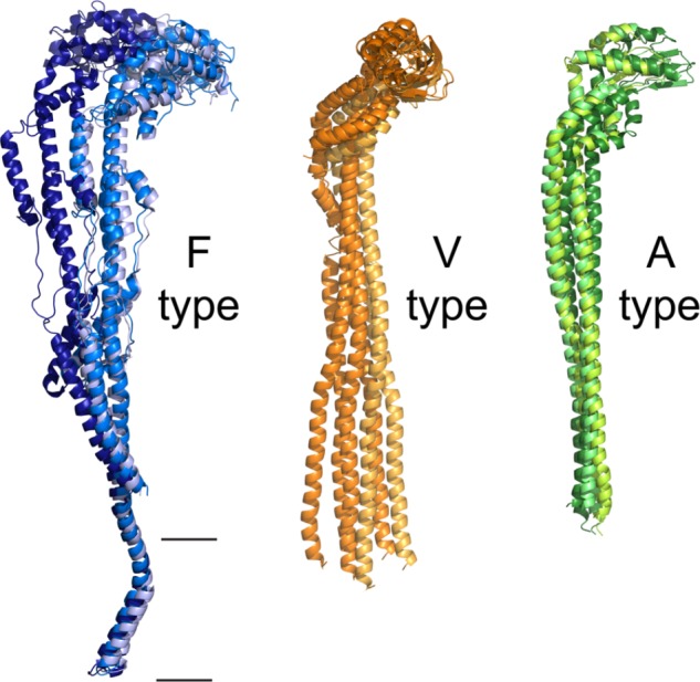
FIGURE 7.

Flexibility of the peripheral stalk of rotary ATPases. Flexibility of the peripheral stalk illustrated with the transitions it goes through during the process of rotational catalysis. (A) Models that correspond to the transitions of an F-ATPase peripheral stalk; PDBs 5ARI, 5ARA, 5FIL from Zhou et al. (2015). The membrane section in these models is not accurately represented due to the resolution of the maps. Higher resolution maps have been obtained but only in one rotational state and thus are not useful to illustrate the flexibility of the peripheral stalk. (B) Models that correspond to three rotational states of one of the peripheral stalks of the V-ATPase of S. cerevisiae; PDBs 3J9T, 3J9U, 3J9V from Zhao et al. (2015). (C) Models that correspond to three rotational states of one of the peripheral stalks of the A-ATPase of T. thermophilus; PDBs 5Y5X, 5Y5Z, 5Y60 from Nakanishi et al. (2018). The black horizontal lines indicate the mitochondrial inner membrane.
