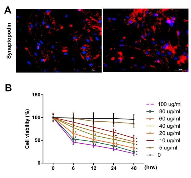
Figure 1.
PAN inhibited the proliferative ability of podocytes in a dose- and time-dependent manner. (A) Synaptopodin expression in podocytes was detected via immunofluorescence assay using a fluorescence microscope (magnification, ×200). (B) Cell viability in podocytes was analyzed using an MTT assay following g treatment with PBS (control), or PAN (5, 10, 20, 40, 60, 80 and 100 µg/ml) for 0, 6, 12, 24 and 48 h time intervals. PAN, puromycin aminonucleoside. *P<0.05 vs. 0 h.

