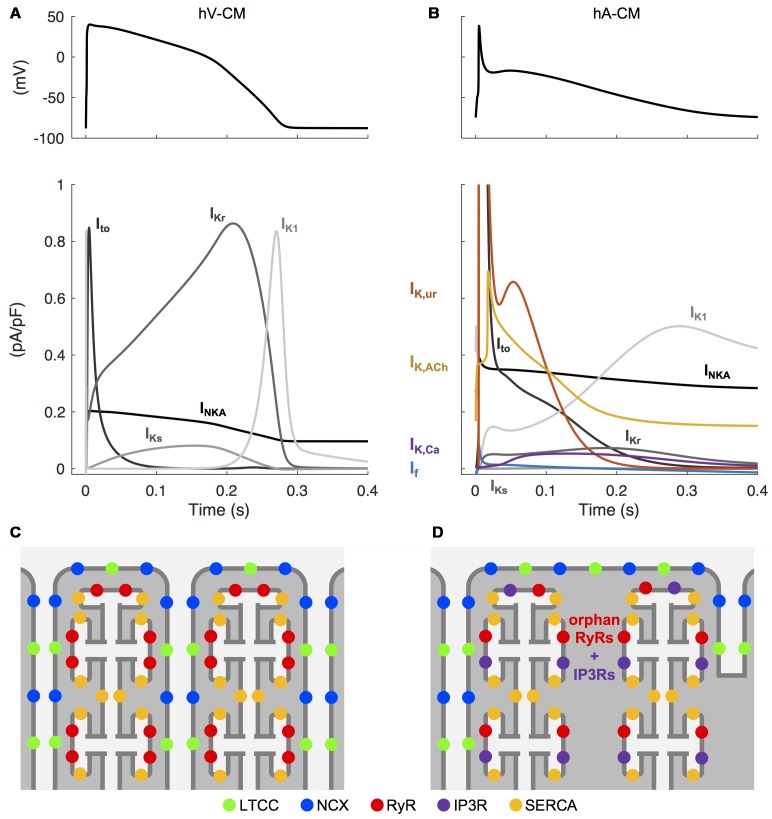
FIGURE 2.
Unique electrophysiological and structural characteristics of atrial cardiomyocytes. Simulated AP in human ventricular (A) and atrial (B) CMs, as well the differences in underlying repolarizing potassium currents, obtained with the hV-CM (O’Hara et al., 2011) and hA-CM (Skibsbye et al., 2016) models, at 1 Hz pacing. Please, note that in order to visualize the weaker currents, the peaks of atrial transient outward potassium current (Ito) (∼11 pA/pF) and the ultra rapidly activating delayed rectifier potassium current (IKur) (∼4.6 pA/pF) are cut off. The atria-specific ion currents funny current (If), IKur, the acetylcholine-activated potassium current (IK,ACh), and the small conductance calcium-activated potassium current (IK,Ca) are highlighted with colored lines. Illustration of the relative positioning of some of the key components involved in intracellular calcium dynamics in hV-CMs (C) and hA-CMs (D).

