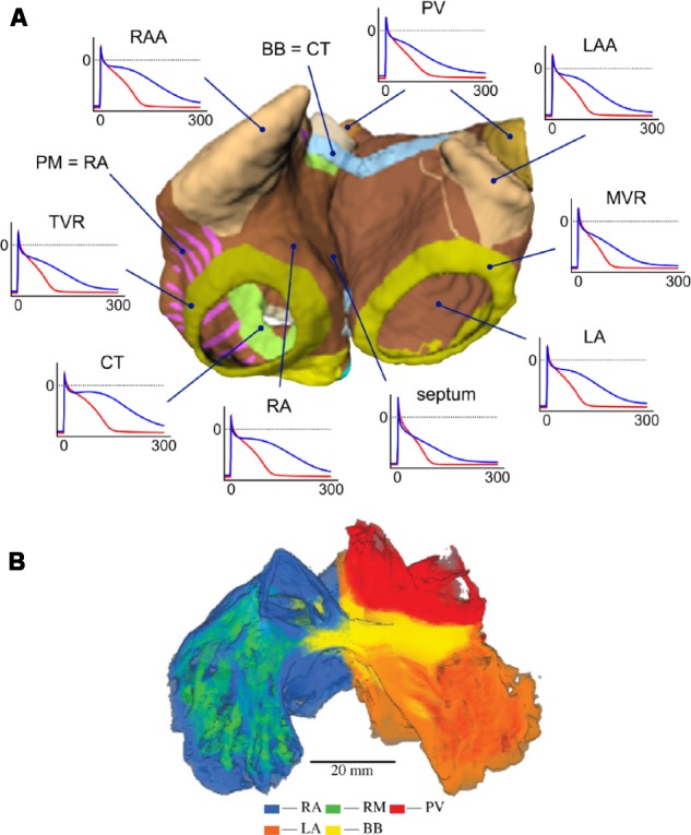
FIGURE 7.

(A) Three-dimensional atrial model with segmented regions and corresponding APs in physiological (blue) and AF-remodeled conditions (red), obtained with the Courtemanche model. From Krueger (2013). Creative Commons Attribution License BY-NC-ND. (B) Regional segmentation of an atrial sheep model into right atrium (RA), left atrium (LA), pectinate muscles (PM), pulmonary veins (PV), and Bachmann’s Bundle (BB). From Butters et al. (2013). Copyright 2013 by Royal Society (United Kingdom). Reprinted with permission.
