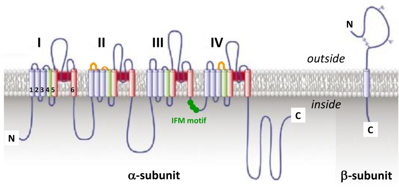
FIGURE 3.
The voltage-gated sodium channel. Schematic representations of α-subunit and auxiliary β-subunit of NaV channels, in which cylinders are transmembrane α helices. In red: S5 and S6 pore-forming segments, in green: S4 voltage-sensor segment, and in blue: S1, S2, and S3 segments. IFM, isoleucine, phenylalanine and methionine residues. The orange loops in DII and DIV domains correspond to spider toxins binding sites (adapted from Catterall et al., 2007).

