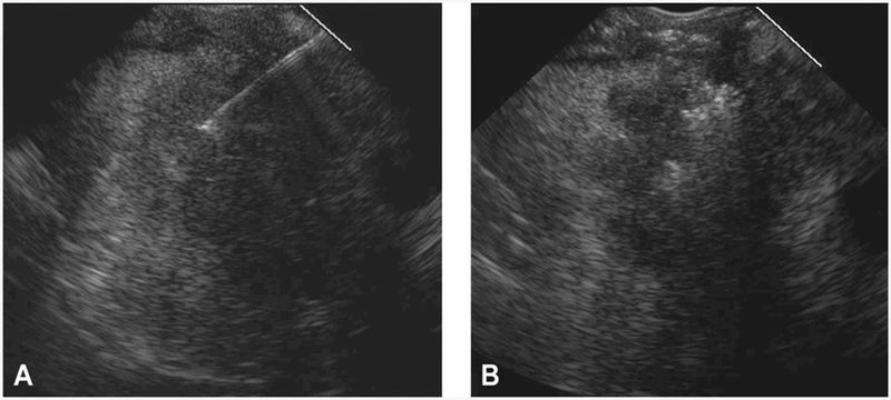
Figure 3.
EUS from a 73-year-old man with PC demonstrating more of a less complete pattern and extent of intratumoral gemcitabine spread assigned a score of 1 out of 4. A, EUS appearance before EUS-FNI reveals a hypoechoic, homogenous mass with an irregular border. B, After EUS-FNI, the hyperechoic-appearing gemcitabine infiltrated only a small portion of the mass and in a nonuniform pattern of spread. The hyperechoic-appearing region is restricted to the central portion of the mass. Most of the gemcitabine extruded from the tumor and can be seen within the peripancreatic space. PC, pancreatic cancer; FNI, fine-needle injection.

