Fig. 8.
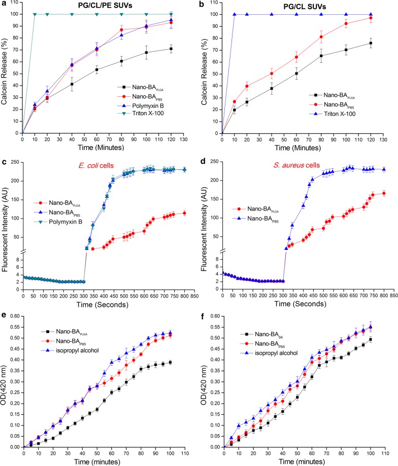
Nano-BAP85-induced calcein release as a function of time. Nano-BAP85 were added to PG/CL/PE SUVs (a) and PG/CL SUVs (b) encapsulated with calcein. Cytoplasmic membrane potential variation of E. coli (c) and S. aureus (d) treated with Nano-BAP85 at 1 × MIC, as assessed by the release of the membrane potential-sensitive dye disC3-5. The fluorescence intensity was monitored at λex = 622 nm and λem = 670 nm as a function of time. Effect of Nano-BAP85 on the cytoplasmic membrane permeability of E. coli cells (e) and S. aureus cells (f). The graphs were derived from average values of three independent trials

