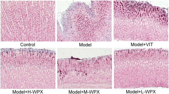
Fig. 2.
Histological evaluation of gastric intestinal metaplasia. Neutral mucins present in normal mucosa were stained red. Sialomucins expressed only in small intestinal-type metaplasia (S-IM) were stained blue, and sulfomucins present in colonic-type metaplasia (C-IM) were stained brown. Images of model gastric epithelium depicted prominent S-IM and C-IM lesions, which were dramatically reduced after WPX administration. n = 9 in each group. (HID-AB-PAS staining, 100×)

