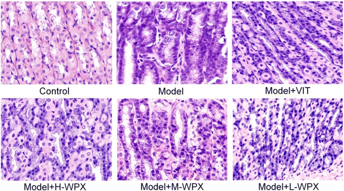
Fig. 3.
Histological evaluation of gastric epithelial dysplasia. Model gastric epithelium displayed GED pathology characterized by glandular architectural abnormalities such as splitting, elongated and crowded glands, back to back formation, as well as by cytological atypia with rounded, pleomorphic nuclei that display prominent nucleoli and loss of polarity. After WPX intervention, these GED pathological alterations, especially irregularities of glandular structure, were regressed in varying degrees. n = 9 in each group. (H&E staining, 100×)

