Fig. 5.
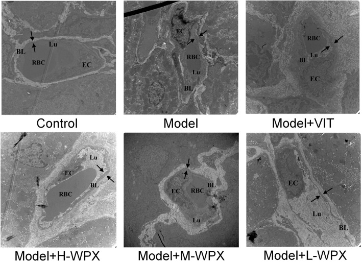
Representative electron micrographs of microvessels in gastric mucosa. Control group: TEM observation of control microvessel ultrastructures appeared intact, in terms of vascular lumen, basal lamina and endothelial cell. Model group: Microvessels lost their typical structures. Vascular lumen, frequently plugged by erythrocytes, was dilated but with a markedly decreased inner diameter. Clearly thickened, rough basal lamina was coated by abundant high-density granules aggregation. Segmental breakup of basal lumina and increased vascular permeability were also existed. Endothelial cells were conglobated and shaped as grapes, characterized by debased cytoplasm electron-density, nucleus chromatin condensation, as well as numerous pinocytotic vesicles. Treatment group: Microvascular abnormalities were still prominent in VIT-treated tissues. However, the abnormalities reversed markedly in WPX-treated tissues, especially in terms of vascular lumen and basal lamina. Even in a few cases, the microvessels were detected to ultrastructurally resemble the normal ones. Note: Opposing arrows mark the thickness of basal lamina; EC, endothelial cell; BL, basal lamina; Lu, lumen; RBC, red blood cell. n = 9 in each group. (10000×)

