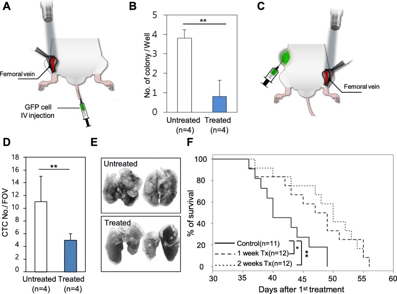
Fig. 2.
CTC-targeting PDT in the GFP-expressing cancer cell-injected mice model and in a syngeneic mice model implanted with GFP-expressing cancer cells. a Irradiation of the mouse femoral vein under the skin flap with a 473-nm wavelength laser after GFP-expressing cancer cell injection via the tail vein. b Clonogenic assay using whole blood taken after the experiment. Colonies were stained with Coomassie blue dye, and the number was compared between each group. Error bar means standard deviation. **, P < 0.01. c Irradiation of the mouse femoral vein under the skin flap with a 473-nm wavelength laser in a syngeneic mouse model with implanted GFP-expressing 4T1 cells. d The number of circulating tumor cells in the 2 weeks treatment and untreated mice. Error bar means standard deviation. FOV means the field of view. **, P < 0.01. e Images of the lungs isolated from mice belonging to the 2 weeks treatment and untreated group. f Kaplan–Meier survival curves of the mice in the control and 1 week treatment and 2 weeks treatment groups. p values were calculated using the log-rank test between treatment groups and control. *, P < 0.05; **, P < 0.01

