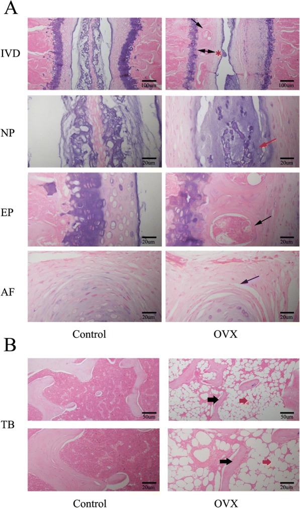
Fig. 4.

Representative images of H&E staining of the intervertebral disc (IVD) and tibia (TB). a Panoramic images of IVD pathology and higher magnification of the endplate (EP), nucleus pulposus (NP), and annulus fibrosus (AF). Ossific nodules (black arrows) in the endplate along with thickening of bony endplate (double arrow) and thinning of the cartilage endplate (red asterisk) were indicated in ovariectomized (OVX) mice. In addition, reduction of notochord cells, degeneration of nucleus pulposus (red arrow), and cleft formation within the annulus fibrosus (blue arrow) appeared in OVX mice. b Images of tibial pathology. Black arrows demonstrate slender trabecular bone and red arrows indicate fat droplets in the bone marrow of OVX mice. n = 9 per group; scale bars = 100 μm, 50 μm, and 20 μm as indicated
