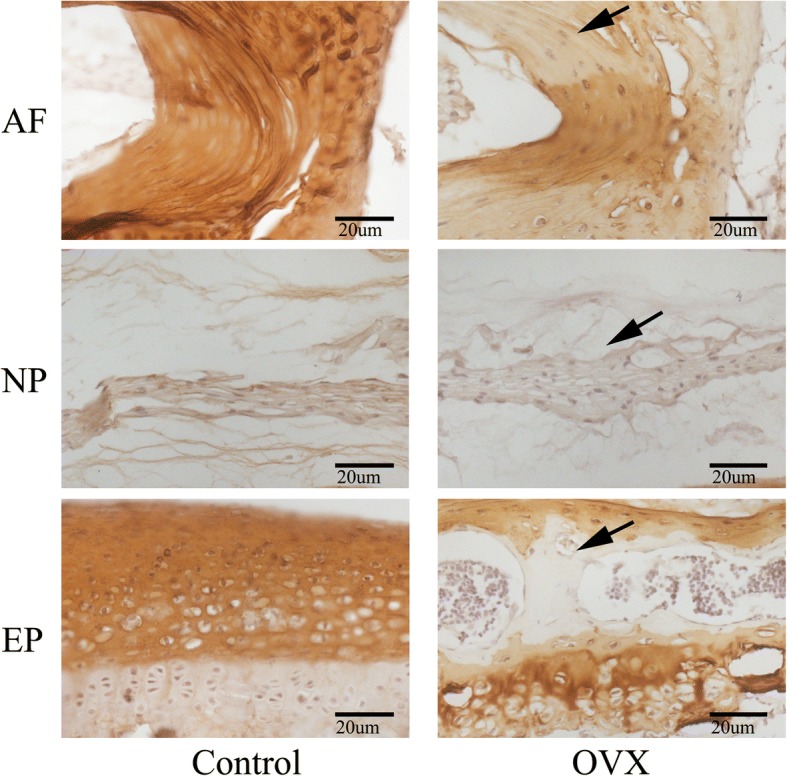
Fig. 8.

Representative sections of immunohistochemistry of Col II. Positive immunostaining was noted as brown staining. Black arrows indicate loss of Col II, especially in the ossification area of the cartilaginous endplate (EP) and the outer layer of the annulus fibrosus (AF) in ovariectomized (OVX) mice. n = 9 per group; scale bars = 20 μm as indicated. NP nucleus pulposus
