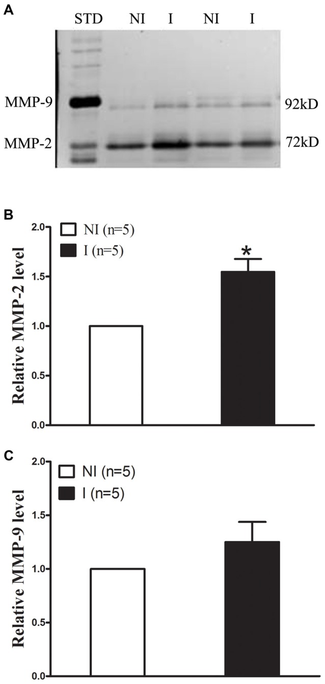
Figure 2.

Effect of 2-h ischemia on MMP-2/9 induction. MMP-2/9 was induced in ischemic brain tissue after 2-h ischemia. (A) Representative gelatin zymogram showed MMP-2/9 levels in the non-ischemic (NI) and ischemic (I) hemispheric tissue. MMP-2 bands were much stronger than MMP-9 bands on zymogram gels. STD is a mixture of standard MMP-2/9. The relative band intensity of MMP-2 (B) and MMP-9 (C) was quantified. A significant increase was observed for MMP-2 in the ischemic tissue after 2-h middle cerebral artery occlusion (MCAO; *P < 0.05 vs. NI, n = 5), while there was no significant increase for MMP-9 (P > 0.05 vs. NI, n = 5). Data were expressed as mean ± SEM.
