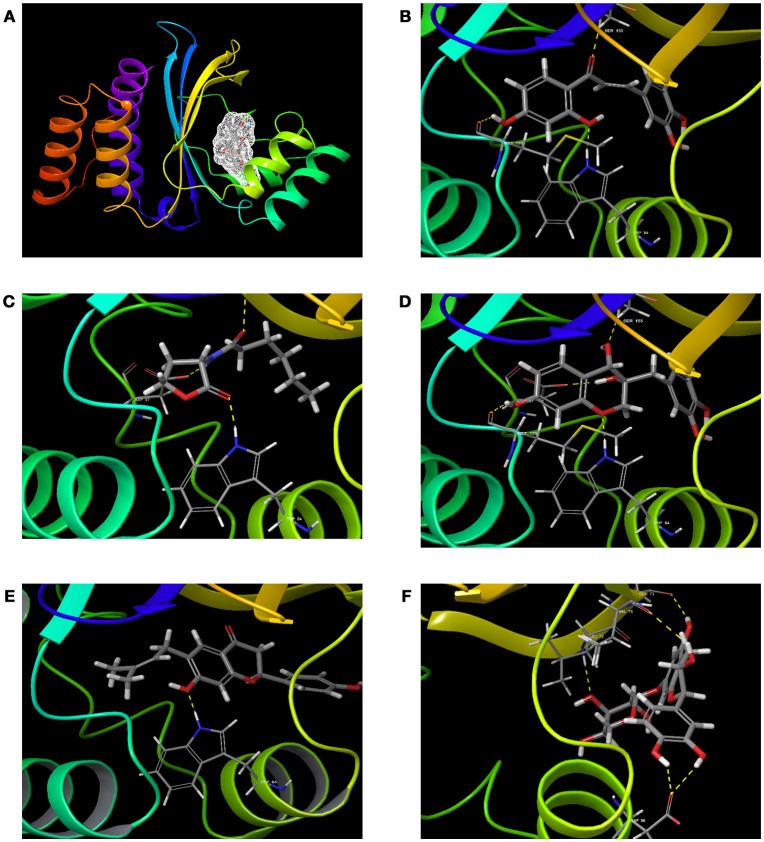
Figure 2.
The 3D molecular interactions of QSIs against CviR (A) is for CviR, the LuxR homolog of C. violaceum where the ligand binding pocket is depicted in light gray (B) is for molecular interaction of C6HSL with CviR (3 H bonds) (C) for molecular interaction of SPL (4 H bonds) (D) for molecular interaction of BN1 (3 H bonds) (E) for molecular interaction of BN2 (1 H bond) and (F) for molecular interaction of C7X against CviR (4 H Bonds). H-bonds represented in yellow dotted line.

