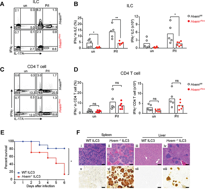
Figure 6. HVEM deficiency affects IFNγ production by ILC during YE infection.

(A,C) Representative plots of IFNγ and IL-17A expression by ILC (CD45+Lin−CD3− CD90.2+)(A) or CD4+ T cells (CD45+Lin−CD3+CD4+)(C). (B,D) Frequencies and absolute numbers of IFNγ expressing ILC (B) and CD4+ T cells (D) from ileal LPL isolated from Hvemfl/fl and HvemᐃRorc mice at day 3 p.i. (2×10⁸ YE CFU/mouse; n=5–6 per group; cohoused littermates). Cells were either unstimulated (un) or stimulated for 4h with P/I and BFA was added in the last 2h of incubation before analysis for intracellular cytokine. (E- F) CCR6− ILC3 (Lin−CD3−NK1.1−CD90.2highCD45intCCR6−) were sorted from SI-LP of Rag1−/− Hvem−/− or Rag1−/− Hvem+/+ mice. Sorted CCR6− ILC3 (~1.5 × 10⁵ cells/mouse) were transferred by retro-orbital injection into Rag2−/−gc−/− recipients at day −1. Groups of mice were infected orally with YE (1.4 × 10⁸ YE CFU/mouse; n=7–11 per group; cohoused). (E) Survival curves. (F) Representative H&E stained (i-iv) or Warthin-Starry silver stained (v-viii) tissues from the indicated mice at day 6 p.i. Images of spleen (i,ii,ν,νi) and liver (iii,iν,νii,viii). Scale bars, 100μm. Two independent experiments were carried out yielding similar results. Statistical analysis was performed using Mann- Whitney test (B,D) or Log-rank test (E). Statistical significance is indicated by*, p < 0.05; **, p < 0.01; ns, not significant. In B and D, bars show the mean and each symbol represents a measurement from a single mouse. Data represent pooled results from two independent experiments having at least three mice per group in each experiment. See also Figure S5.
