
Figure 1: Development of a multiplexed imaging assay for the tumor-immune microenvironment.
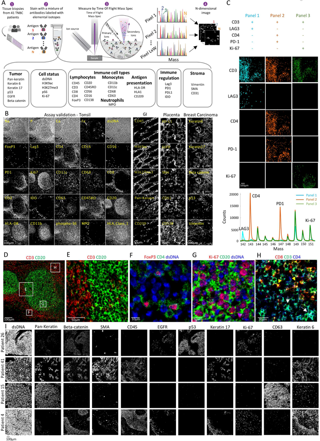
(A) Experimental workflow for MIBI-TOF. Clinical tissue specimens are stained using a mixture of antibodies labeled with elemental mass tags. The samples are rasterized by a primary ion beam that releases lanthanide adducts of the bound antibodies as secondary ions, which are recorded by a TOFMS. This results in an n-dimensional image depicting protein expression in the field. (B) MIBI-TOF images of samples stained with the panel in (A). (C) Serial sections of lymph node were stained with three sub-panels, each including 30 out of the 36 antibodies. Bottom: integrated counts across the image as a function of mass for the three panels. (D-H) Assay validation: Color overlays of tonsil show expected patterns of histologic architecture, subcellular staining (membrane/nucleus), and protein co-expression. (I) MIBI-TOF images of four TNBC patients.