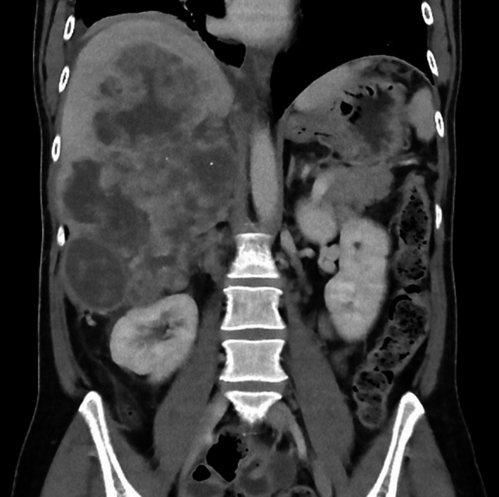
Figure 1.

Abdominal CT scan. An axial contrast‐enhanced computed tomography (CT) image of the abdomen shows an inhomogeneous, large, nonenhanced hypodense lesion measuring 13.6 × 11.6 × 20 cm, occupying most of the right liver with exophytic components encroaching the upper right suprarenal region and displacing the right kidney inferiorly
