
Figure 4.
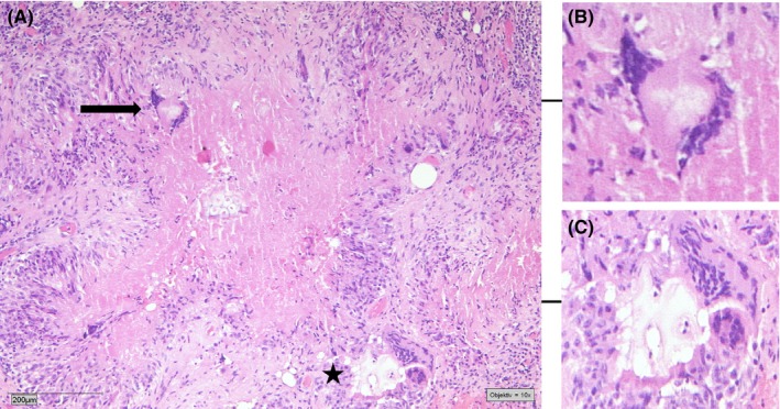
Histological finding of the peritoneal biopsies, showing focal fibrosis areas with local hemorrhages and epithelioid granulomas with central necrotic regions (A). Some giant cells contain phagocytosed foreign material (black arrow, B). The granuloma is surrounded by a diffuse inflammatory cell infiltration, consisting of lymphocytes, neutrophils, plasma cells, macrophages, and some multinucleate giant cells surrounding foreign material (black star, C). (Hematoxylin and Eosin staining, 10× magnification)