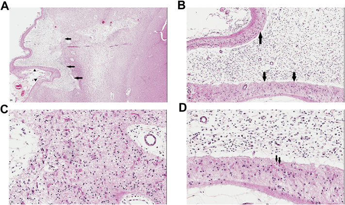
Fig. 2.

A. Remote or ‘old’ cystic infarct in cortex lateral to the basal ganglia (visible at right of the micrograph; all images are different views and magnifications of the same infarct). Arrows indicate a cystic cavity representing the infarct. Arrowheads indicate preserved layer I of the cortex, a characteristic finding in old ‘strokes’. B. Magnified view of the infarct shows the cystic cavity replete with lipid-laden macrophages; arrows indicate the preserved and clearly outlined layer I of the cortex, immediately underlying leptomeninges. C. Deep ‘margin’ of the cystic infarct shows abundant astrocytes, including gemistocytes, with prominent eosinophilic cytoplasm. D. Magnified view of the interface between preserved cortical layer I (bottom of the image) and lipid laden macrophages (at top of the image). Subarachnoid space is at the bottom edge of the micrograph. Arrows indicate two gemistocytes with prominent cell processes. [All images are from sections stained with hematoxylin and eosin/H&E].
