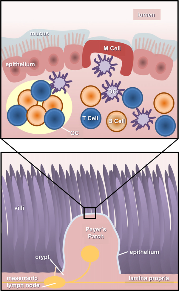
Fig. 2.
Anatomy of the gastrointestinal immune system. There are two main components in the gut-associated lymphoid tissue (GALT): inductive and effector sites. Inductive tissues include the action of Peyer’s patches, lymphoid follicles (within lymph nodes), and antigen presenting cells (APCs). Meanwhile, effector sites comprise the lamina propria and the surface epithelium. Upon entering to the intestinal lumen, antigens are transported across the intestinal epithelium barrier by sampling M cells, transcytosed and delivered to APCs (i.e. DCs). Activated DCs travel and prime CD4+ T cells in germinal centers (GCs), present in the Peyer’s Patches (PPs) and mesenteric lymph nodes, needed to initiate an immune response. Primed CD4+ T cells then activate B cells, which undergo isotype switching, thus generating IgA+ B cells. These B cells then leave the PPs using the lymphatic system to enter circulation and reach effector sites in the lamina propria, mature, and become IgA producing-plasma B cells.

