
Figure 2.
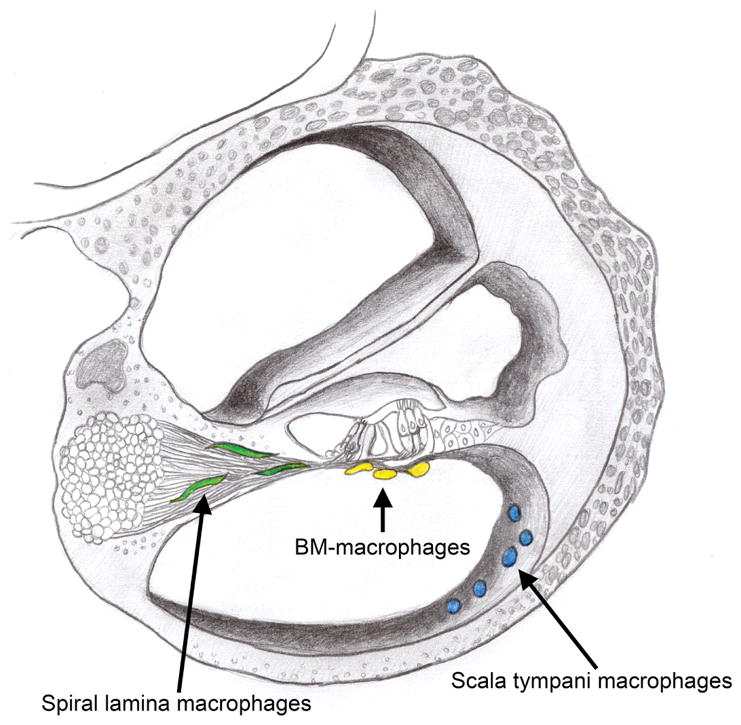
Schematic drawing of a cochlear cross-section indicating the location of cochlear macrophages in three anatomic sites. The osseous spiral lamina macrophages reside closely among the peripheral fibers of the spiral ganglia and are color-coded green in this schematic. BM-macrophages inhabit the scala tympani cavity and are found immediately beneath the basilar membrane in close proximity to the sensory cells and their innervations by spiral ganglia (color-coded yellow). Cochlear macrophages on the luminal surface of the scala tympani cavity were also examined and are color-coded blue.