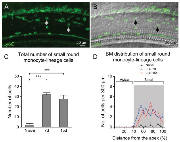
Figure 6.
Analysis of macrophages with a small round shape beneath the basilar membrane following LLN. A. A typical image of Ly6C-positive cells (pointed to by arrows) beneath the basilar membrane in the middle turn of a cochlea following LLN for 15 days. B. DIC view of the same cochlear site. C. The total number of small round Ly6C-positive cells increases following exposure to LLN (n=5 cochleae for each time point) (*** indicates P < 0.001). D. Distribution of Ly6C-positive cells along the basilar membrane. Note that the increase in the number of Ly6C-positive cells is specifically found within the middle to basal portion (40–100% from apex) of the basilar membrane of cochleae stressed by LLN. This cell type was comparatively scant in all basilar membrane portions of naive cochleae (data not shown).

