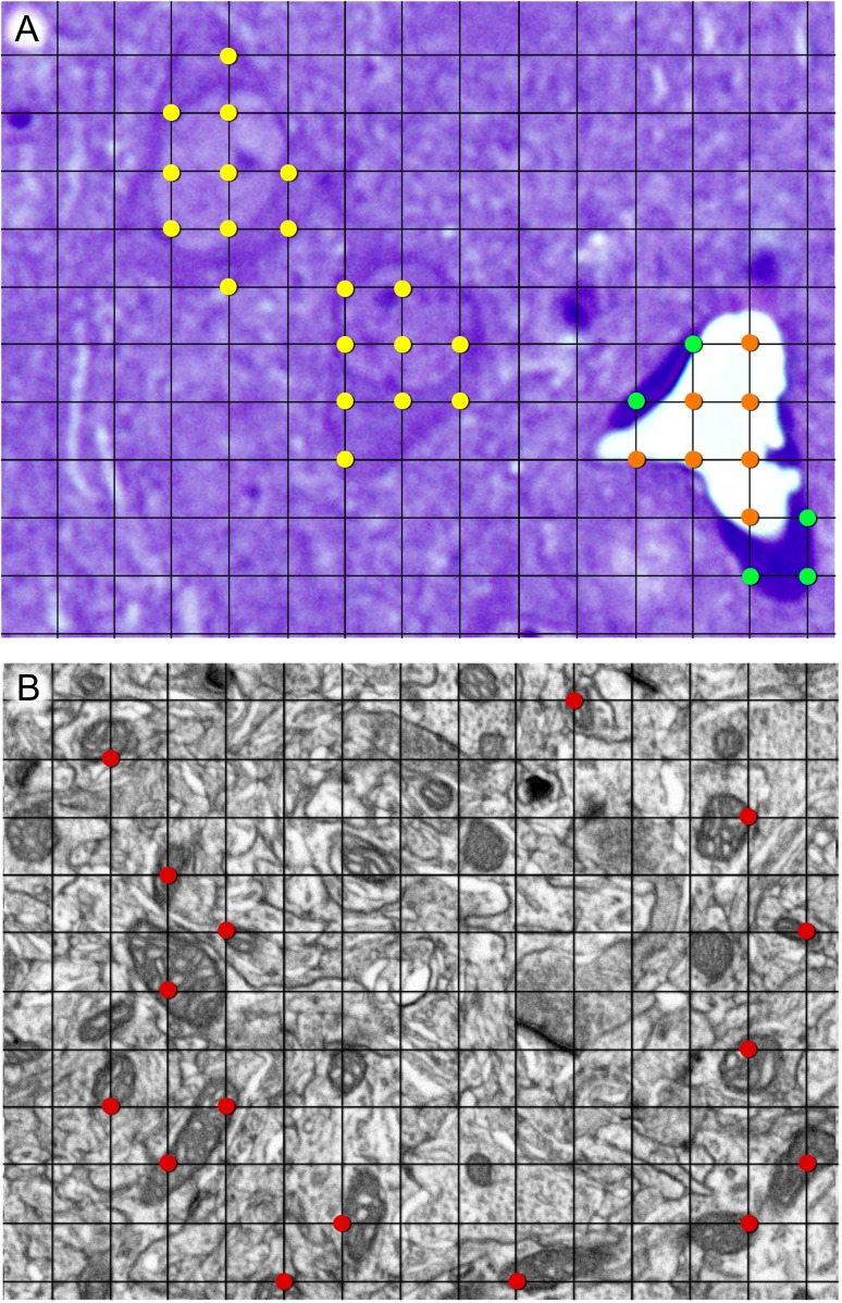
Figure 1.
Stereological grids used for the estimation of the volume fraction of different compartments at the light (A) and electron microscopic (B) levels, using the Cavalieri method. (A) Example of the estimation of the volume fraction of neuropil, cell somata, and blood vessels at the light microscopic level. Points hitting cell somata, blood vessel lumen, and blood vessel endothelium have been highlighted in yellow, orange, and green, respectively. The remaining points hit the neuropil, although they have not been highlighted for the sake of clarity. (B) Example of one stereological grid used for the estimation of the volume fraction of mitochondria at the electron microscopic level. Points hitting mitochondria have been highlighted in red. The ratio of grid points hitting any compartment to points hitting the reference area is proportional to the volume fraction occupied by that compartment (see Materials and Methods for details). The thickness of grid lines and the size of marked points have been exaggerated for clarity. Grid size is 4 × 4 μm2 in (A) and 500 × 500 nm2 in (B).

