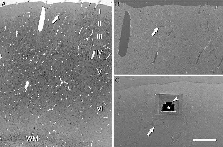
Figure 2.
An example of the use of a semithin section to locate the region of interest. (A) Semithin section of the somatosensory cortex stained with toluidine blue and photographed with an optical microscope. This section is immediately adjacent to the block face that was later photographed with the SEM. Cortical layers (I–VI) and white matter (WM) can be identified. Blood vessel profiles can be used as reference points to locate the region of interest. In this particular example, a vascular profile (arrow) is located approximately in the boundary between layers I and II. (B) Low power electron micrograph acquired with the SEM from the block surface. The same vascular profile that was previously identified in the semithin section is also visible on the block face (arrow). (C) The same block after a series of images has been acquired with the FIB/SEM. The reference blood vessel (arrow) was used to locate layer I. First, a large trench (asterisk) was excavated with the FIB in order to have visual access to the tissue below the block surface. Then, a smaller trench (arrow head) was sequentially milled with the FIB and photographed with the SEM at intervals of 20 nm, thus obtaining a series of images. A similar procedure was used to locate the regions of interest in all other cortical layers. Scale bar = 300, 150, and 80 μm in (A), (B), and (C), respectively. See also Supplementary Video 1.

