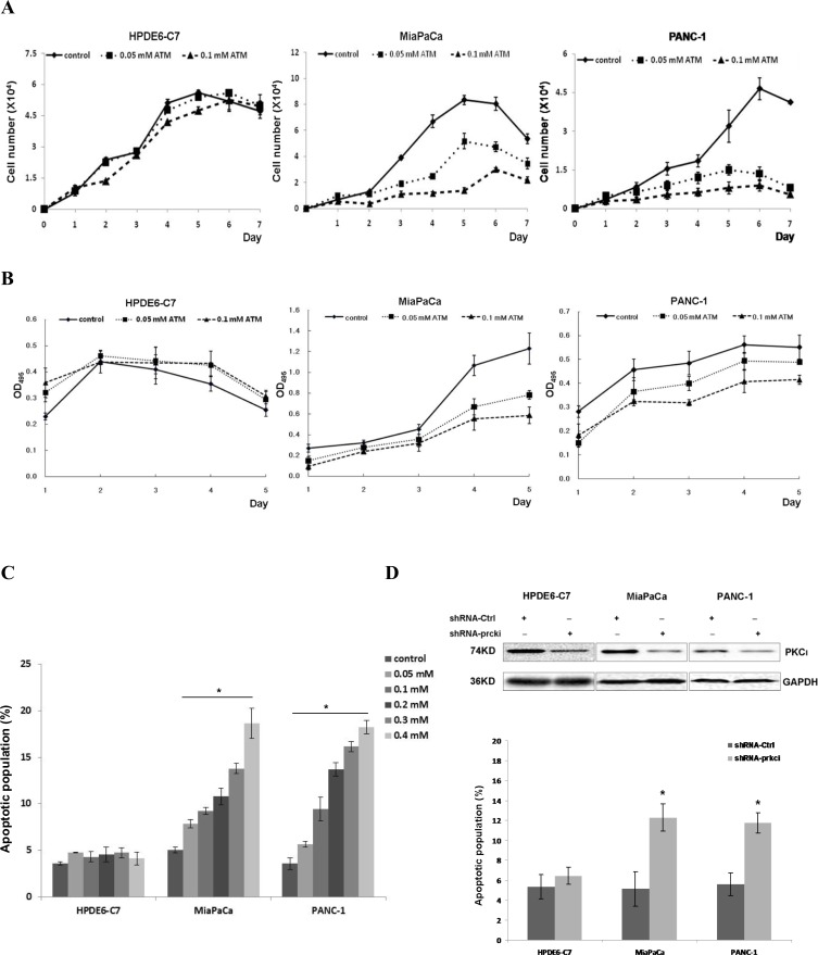
Figure 1. PKCι inhibition suppresses the growth and induces apoptosis of pancreatic cancer MiaPaCa and PANC-1 cells harboring aberrant Kras, while showing little effect on non-neoplastic pancreatic epithelial HPDE6-C7 cells containing normal Kras.
(A) Representative growth curves of HPDE6-C7, MiaPaCa and PANC-1 in presence of control diluent, 0.05 or 0.1 mM ATM. Cells were counted in triplicate wells at each time point and the result represents three independent experiments (p < 0.05). (B) Proliferation of HPDE6-C7, MiaPaCa or PANC-1 treated with control diluent, 0.05 or 0.1 mM ATM as determined by MTT colorimetric assay. Error bars represent SD of 3 replicates samples (p < 0.05). (C) Quantification of apoptotic cells induced by increasing concentrations of ATM in HPDE6-C7, MiaPaCa and PANC-1. The experiments were repeated at least five times (Error bars, ± SD. n = 5 per time point. *p < 0.05). (D) Upper panels: Immunoblot analysis of PKCι expression in HPDE6-C7, MiaPaCa and PANC-1 infected with lentivirus producing non-targeting scrambled shRNA (shRNA-Ctrl) or shRNA targeting human PKCι (shRNA-prkci). Lower panel: Quantification of apoptotic cells in HPDE6-C7, MiaPaCa and PANC-1 expressing lentiviral shRNA-Ctrl or shRNA-prkci. Data represent mean of 5 replicates ± SD. *p < 0.05.

