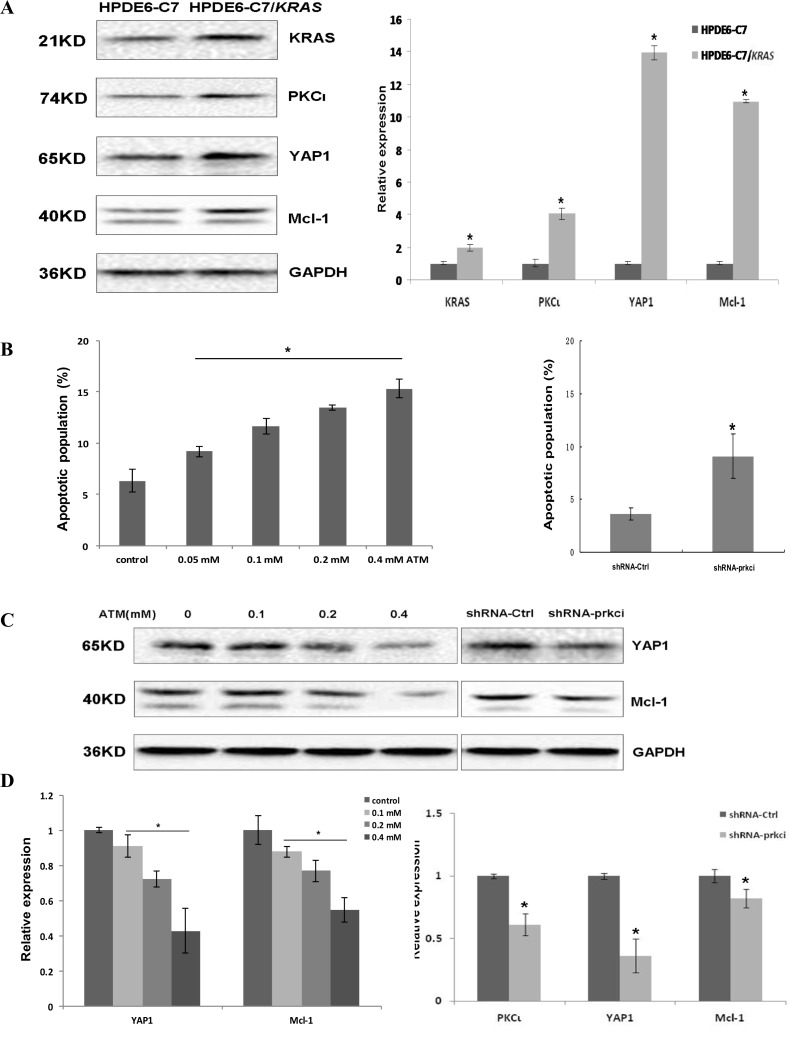
Figure 7. Stable transfection of HPDE6-C7 with KRASG12C renders the cells sensitivity towards PKCι inhibition.
(A) Immunoblot (left panels) and qRT-PCR (right panel) analyses demonstrating the upregulation of Kras, PKCι, YAP1 and Mcl-1 in HPDE6-C7/KRAS in comparison with its parental HPDE6-C7 cell line(Error bar, ± SD. n = 3. *p < 0.05). (B) Quantification of apoptotic cells in HPDE6-C7/KRAS treated with increasing concentrations of ATM (left panel), and cells expressing lentiviral shRNA-Ctrl or shRNA-prkci (right panel. Error bar, ± SD. n = 5. *p < 0.05). (C) Immunoblot and (D) qRT-PCR analyses of YAP1 and Mcl-1 expression in HPDE6-C7/KRAS treated with increasing concentrations of ATM, or infected with lentivirus expressing shRNA-Ctrl or shRNA-prkci (Error bar, ± SD. n = 3. *p < 0.05).

