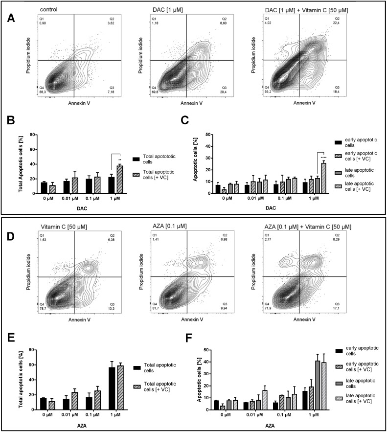
Figure 9. Increase of apoptotic cells in human colon cancer cells by vitamin C and DAC/AZA incubation.
Representative contour plots of Annexin V/propidium iodide staining in HCT116 colon cancer cells treated for 72 h with DAC (A) and vitamin C, alone or in combinations, as indicated. Q1 gates for necrotic cells, Q2 for late apoptosis, Q3 for early apoptosis and Q4 for healthy cells. TNFα (20 ng/mL) and cycloheximide (20 μg/mL) were used as positive controls for the induction of apoptosis. Quantitative representation of total apoptotic cells (B) and early + late apoptotic cells separately (C) after treatment with DAC with or without vitamin C, with indicated concentrations. Representative contour plots of Annexin V/propidium iodide staining in HCT116 colon cancer cells treated for 72 h with AZA in indicated concentrations and vitamin C [50 μM] (D). Quantitative representation of total apoptotic cells (E) and early + late apoptotic cells separately (F) after treatment with AZA with or without vitamin C, with indicated concentrations (error bars = SD; n=3; Statistical significance of the treated groups to the untreated control was calculated using 2-way ANOVA and Tukey post-test **** = p< 0.0001, *** = p<0.001, ** = p<0.01, * = p<0.05).

