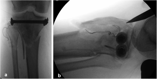
Fig. 4.

Figs. 4-A and 4-B Intraoperative anteroposterior (Fig. 4-A) and lateral (Fig. 4-B) radiographs made after the insertion of 2 compression bolts. The lateral radiograph also shows the entry point for the intramedullary nail. The fractured anterior metaphyseal area was treated with moderate release of the calcaneal traction and 2 anteroposterior free lag screws at a later stage as shown in Figure 5.
