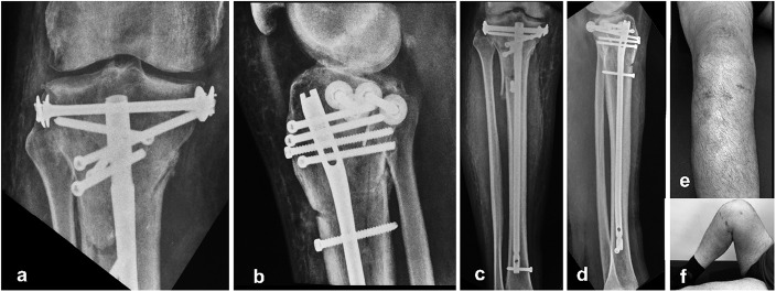
Fig. 6.
Figs. 6-A through 6-F Images made at the time of the latest follow-up. Figs. 6-A and 6-B Anteroposterior (Fig. 6-A) and lateral (Fig. 6-B) radiographs of the knee joint. Figs. 6-C and 6-D Anteroposterior (Fig. 6-C) and lateral (Fig. 6-D) radiographs showing the entire tibia. Figs. 6-E and 6-F Photographs of the lower extremity, showing the restoration of the anatomy and the function of the knee joint as well as the minimal invasiveness of the technique.

