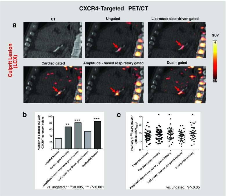
Fig. 5.
Effect of motion correction on [68Ga]pentixafor uptake and detectability in culprit coronary lesions. a [68Ga]Pentixafor PET/CT images of a culprit LCX lesion. Focal CXCR4 signal (red) is more clearly depicted after cardiac, respiratory motion or dual cardiac/respiratory motion correction (lower row) compared to ungated images (upper row, middle). b Detection rates for CXCR4+ culprit coronary lesions are higher in gated images (P ≤ 0.005). c Signal intensity of CXCR4+ culprit coronary lesions is higher in gated images (P < 0.05). Background subtraction was performed using an individually adjusted threshold in this particular subject for clearer visualization of tracer uptake

