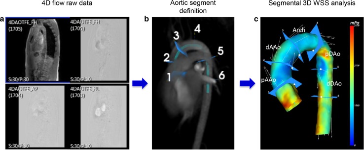
Fig. 1.
Aortic 4D flow MRI processing and analysis. a 4D flow MRI raw data including anatomical and flow data. b Automatic segmentation of 3D aortic volume after manually defining start and endpoint of the thoracic aorta and aortic segment definition. c 3D color-coded aortic segmentation representing WSSmax distribution for one systolic cardiac phase

