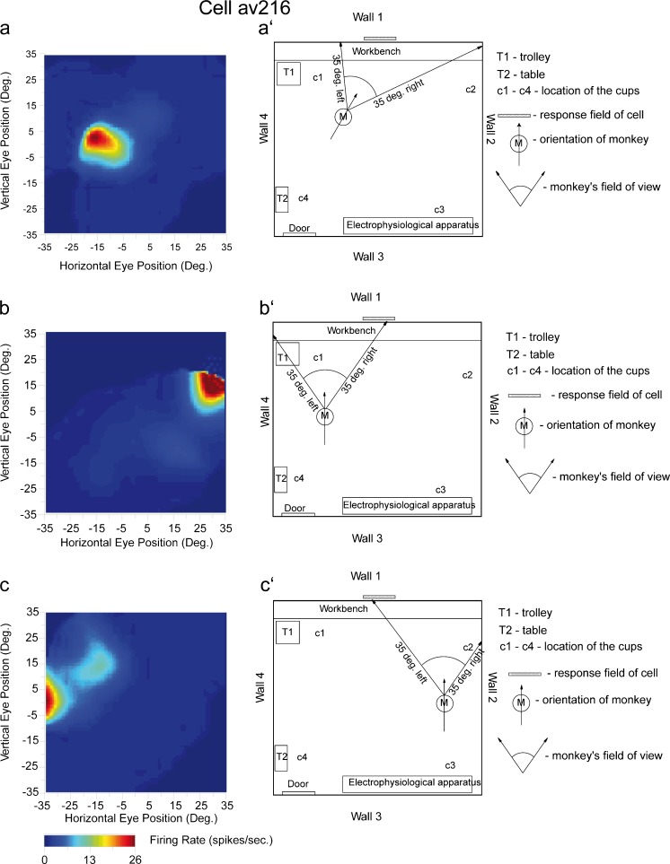
Fig. 3.
Examples of the firing of a hippocampal spatial view cell (av216) when the monkey was at various positions in the room, with various head directions, looking at wall 1 of the room. The details of the spatial view field are shown by the different firing rates with the colour calibration bar shown below. The firing rate of the cell in spikes/s as a function of horizontal and vertical eye position is indicated by the colour in each diagram left (with the calibration bar in spikes/s shown below). Positive values of eye position represent right in the horizontal plane and up in the vertical plane (hatched box right approximate position of spatial view field). The diagram provides evidence that the spatial view field is in allocentric room-based coordinates and not eye position or place coordinates (for details see Georges-François et al. 1999)

