Fig. 4.
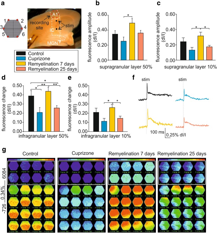
Demyelination alters neuronal network properties in the mouse thalamocortical auditory pathway in vitro. a Micrograph shows the preserved auditory thalamocortical pathway in an acute brain slice including MGN, IC, and A1. Electrical stimulation (stim) was performed at short and long distances (black dots) from A1 and simultaneous electrical and optical recordings were performed. Note the schematic representation of the VSD photodiode array (red hexagon, a). b, c Bar graphs showing the fractional fluorescence change in response to electrical stimulation of 50% (b) and 10% (c) intensity for all experimental groups in the supragranular layer. d, e Bar graphs showing the fractional fluorescence changes in response to electrical stimulation of 50% (d) and 10% (e) intensity for all experimental groups in the infragranular layer. f Example traces showing single exemplary diode traces for all experimental groups in response to 50% SI. g Fluorescence maps representing the spatiotemporal propagation of the VSD signal in A1 in response to electrical stimulation (50%). All experimental groups were normalized to control to appreciate the differences in fluorescence intensity, as shown by the raw diode example traces (control = 0.34% of fluorescence change)

