Fig. 5.
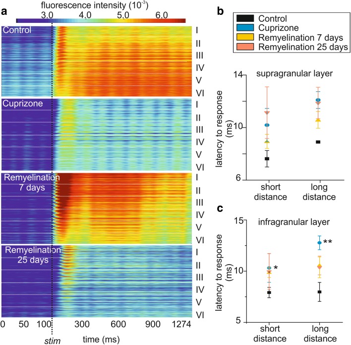
Demyelination altered the latency to response in all cortical layers. a Schematic representation of the spatiotemporal propagation of the evoked stimulus (x-axis = time in ms, y-axis = A1 layers) in response to electrical stimulation which is indicated by the vertical dashed line. b, c Response latency to electric stimulation depends on the position of the stimulation electrode: short and long distances from the cortical recording site (n = 6 and n = 7, respectively) in the supra (b) and infragranular layer (c). The latency significantly increased in cuprizone-treated animals and it is not restored during the early and late remyelination phase. *p < 0.05, **p < 0.01

