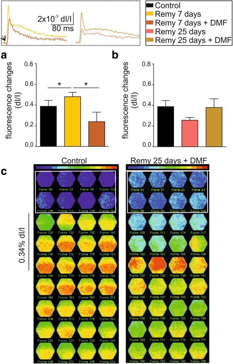
Fig. 6.
DMF treatment improves neuronal network functionality in vitro. a Bar graphs showing the fractional fluorescence changes in response to electrical stimulation of 50% intensity in control animals (black bars) and after 7 days of remyelination with and without DMF treatment (dark orange and yellow bars, respectively). The two left superimposed exemplary traces in the inset show the difference in amplitude between these two groups. b Bar graphs showing the fractional fluorescence changes in response to electrical stimulation of 50% intensity in control animals (black bars) and after 25 days of remyelination with and without DMF treatment (ocher and pink, respectively). The two right superimposed exemplary traces in the inset show the difference in amplitude between these two groups. c Fluorescence maps showing the spatiotemporal pattern of stimulus propagation in control 25 day DMF-treated animals. *p < 0.05

